- Text Size:


9 March 2016
EOS and the University of Michigan customise a biocompatible material for additively manufacturing medical implants
 An adolescent girl has received novel, 3D-printed tracheal splints to treat a congenital breathing condition called tracheobronchomalasia. She joins three baby boys and a baby girl who previously underwent similar groundbreaking operations. All five patients continue to thrive, as their collapsed airways now function normally thanks to the surgical procedure.
It was pioneered by Dr Glenn Green, a paediatric otolaryngologist and his surgical team from CS Mott Children's Hospital, Ann Arbor, who joined forces with Dr Scott Hollister, professor of biomedical engineering and lead researcher at the University of Michigan. Dr Green can be seen explaining the project in a video on YouTube.
Dr Hollister, who first learned about additive manufacturing in the 1990s, said, “Even if a market is relatively small, it doesn’t diminish the human need to be treated. It is estimated that one in every 2,000 children worldwide is affected by this life-threatening condition.
“When I started designing my own porous scaffolds for anatomic reconstruction, I realised that 3D-printing would be ideal for creating the complex geometries I had in mind.
“It is now pretty automatic to generate an individualised splint design and print it; the whole process only takes about two days now instead of three to five”.
Polycaprolactone (PCL) was found to be the perfect material for additively manufacturing a tracheal implant. First, it has a long resorption time, which is very important in airway applications as the implant needs to remain in place for at least two years before it is resorbed. Second, PCL is very ductile, so if it fails it will not produce any particles that might puncture tissue. Third, PCL can be readily processed for and fabricated on an EOS additive manufacturing system.
Dr Hollister and the University of Michigan purchased an EOS FORMIGA P 100 in 2006 to aid research into scaffolds and biomaterials. He continued, “I chose EOS because we were looking for a system that was flexible and allowed us to change parameter settings such as laser power, speed, powder bed temperature and so on, which we needed to do to customise our builds.
“Also, because biomaterials can be expensive and implants and scaffolds are typically not so big, we wanted a more limited build volume that didn’t use a lot of material. The FORMIGA P 100 fitted the bill for both of these requirements.
“EOS even gave us access to software patches to enable us to change the range of parameters of the machine to best process the PCL material.”
Additive manufacturing expertise was provided on site by EOS, who helped the team in their laboratory, advising how best to prepare the material for production. While the company offers a wide variety of proprietary plastic and metal materials, the use of PCL was a first in this case.
Martin Bullemer, Business Development Manager Medical at EOS said, “We make a point of being very open in terms of materials. We support universities in the development of their own parameters for processing novel materials in EOS systems, such as in this instance.”
The university team uses patient data from MRI or CT scans to examine the defect to be repaired, then creates computer models of the anatomy. Engineers are able to use a CAD system to design splints with a highly compliant, porous structure of interconnected spaces that will slowly expand with the maturing airway. Finally, the splint is produced in the EOS FORMIGA P 100 system. After fabrication, researchers measure the splint dimensions and mechanically test them.
Following the operation, the splint-supported trachea expands and functions immediately, so when patients are weaned off oxygen they are able to breathe normally. Kaiba Gionfriddo, the first baby to benefit from the procedure, is now nearly four years old. The boy’s own tissues have successfully taken over the job of the implant, which has been almost completely reabsorbed into his body.
To treat patients with birth defects or following illnesses or accidents, Dr Hollister’s group is also developing craniofacial, spine, long bone, ear and nose scaffolds and implants and additively manufacturing them using a material with characteristics that promote reconstruction and regrowth.
He concluded, “I see a time soon, probably within the next five years, when many hospitals and medical centres will print their own devices specifically for their own patients and not need to get them off the shelf.
“If we can expand the number of biomaterials used in additive manufacturing, we can tackle a tremendous number of problems in all fields of reconstructive surgery and make enormous strides for the benefit of patients.”
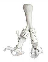
Image: two of the University of Michigan's 3D-printed tracheal splints, made of polycaprolactone using EOS technology, on a model of a trachea.
- Contact Information
- Name: Stuart Jackson
- Email: stuart.jackson@eos.info
- Website: www.eos.info